”分子生物学”の部屋 へようこそ!
ここでは私が、現在行っている研究の最新情報をお届けいたします。
Genomic Sequence Analysis of the Bovine Male-Enhanced Antigen-1 (Mea-1) and Differential Localization of its Transcripts and Products During Spermatogenesis
Kondo M., Terouchi S., Tsukasa N., Sato S., Ishida N. and Sutou S. DNA Sequence, 6, 75-85 (1996)
ABSTRACT
The male-enhanced antigen-2 (Mea-2) gene was originally identified with a monoclonal histocompatibility Y (H-Y) antibody (mAb4VII). There is no report of the full length cDNA encode for Mea-2 product until this report. In this study, we isolated the full length mouse Mea-2 cDNA by screening a testis cDNA library with a PCR-amplified Mea-2 product, and direct PCR amplification of its upstream sequences from the cDNA library. The primary structure of the Mea-2 peptide, deduced from this nucleotide sequence, shows that it encode a 150 kDa protein, of 1325 amino acid residues, which contained five putative N-glycosylation sites and four leucine zipper motifs. A data bank search indicated that it has high homology with a human Golgi autoantigen (golgin-160) both in its nucleotides (78%) and amino acids sequence (83%). This suggests that Mea-2 gene product may encode a golgi structural protein. In situ hybridization analysis suggested that the Mea-2 gene is expressed in spermatids during spermatogenesis as already shown by Mea-1, suggesting that Mea-2 gene product as well as Mea-1 have also some role for spermatogenesis.
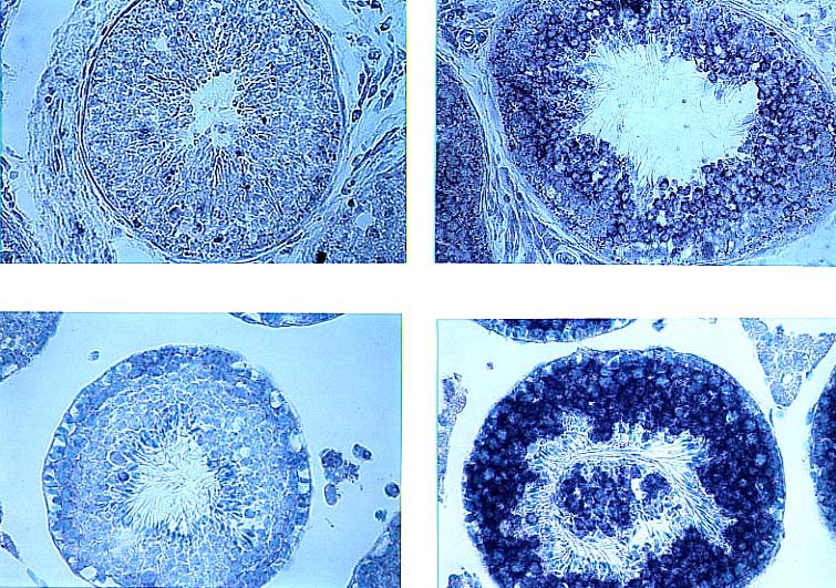 In situ hybridization of the Mea-1 mRNA in the bovine and mouse testis. A, C: In situ hybridization shows the digoxigenin-labeled anti-sense probe of bovine (A) (EcoRI-BamHI 350bp fragment) (Kondo et al., 1993) or mouse (C) Mea-1 cDNA (EcoRI-BamHI 350bp fragment) (Lau et al., 1989) detected specific mRNA signals (wine red) in primary and secondary spermatocytes and spermatids. B, D: The sense Mea-1 probe did not show any signal in bovine (B) or mouse (D) testis (control). Counter-staining for the nuclei was performed with methylgreen (original magnification, 50x).
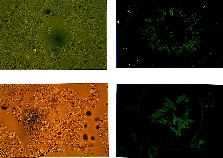 Immunofluorescence localization of Mea-1 product in mouse testis sections. Bovine (4.5 years old) (A, B) and mouse (10 weeks old) testis (C, D) sections were fixed and stained as described in Materials and Methods. Fluorescence was detected specifically only in spermatids. A, C: Phase contrast image; B, D: Fluorescence image of a seminiferous tubule (original magnification, 66x).
Cloning and molecular characterization of cDNA encoding a mouse male-enhanced antigen-2 (Mea-2): A putative family of the Golgi autoantigen
Kondo M. and Sutou S. DNA Sequence, 7, 71-82 (1997)
ABSTRACT
The male-enhanced antigen-2 (Mea-2) gene was originally identified with a monoclonal histocompatibility Y (H-Y) antibody (mAb4VII). There is no report of the full length cDNA encode for Mea-2 product until this report. In this study, we isolated the full length mouse Mea-2 cDNA by screening a testis cDNA library with a PCR-amplified Mea-2 product, and direct PCR amplification of its upstream sequences from the cDNA library. The primary structure of the Mea-2 peptide, deduced from this nucleotide sequence, shows that it encode a 150 kDa protein, of 1325 amino acid residues, which contained five putative N-glycosylation sites and four leucine zipper motifs. A data bank search indicated that it has high homology with a human Golgi autoantigen (golgin-160) both in its nucleotides (78%) and amino acids sequence (83%). This suggests that Mea-2 gene product may encode a golgi structural protein. In situ hybridization analysis suggested that the Mea-2 gene is expressed in spermatids during spermatogenesis as already shown by Mea-1, suggesting that Mea-2 gene product as well as Mea-1 have also some role for spermatogenesis.
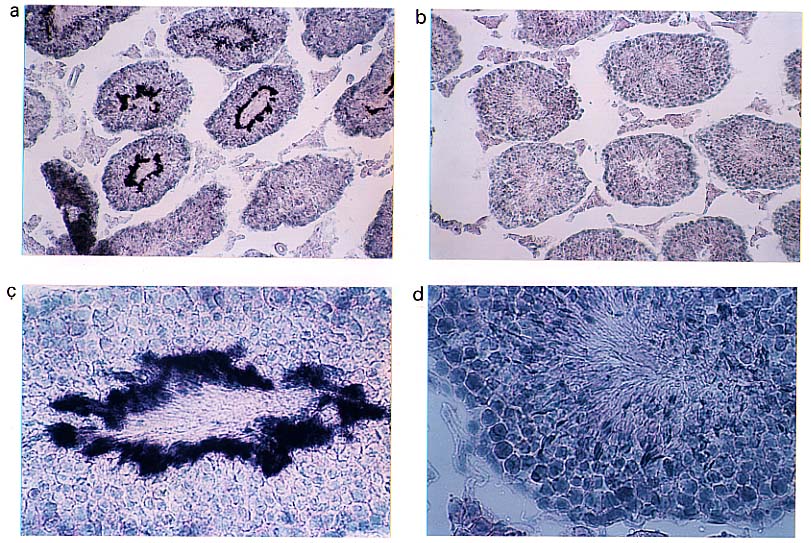 In situ hybridization of Mea-2 mRNA from mouse testes. (a): In situ hybridization shows the digoxigenin-labeled anti-sense probe of Mea-2 cDNA (SphI 456bp fragment, 1081 to 1537 in Fig. 4a) detected specific mRNA signals (wine red) in spermatids. (b): The sense Mea-2 probe did not give any signals in mouse testes. (c), (d): Higher magnification of the seminiferous tuble seen in (a) and (b), respectively. Counter-staining for the nuclei was performed with methylgreen.
Please send your thoughts, suggestions, and discussions to me.
 Go Home
Presented by Masaaki kondo